Image: ShutterStock
The female reproductive system is responsible for producing gametes called ovum or egg (1). Various organ systems in the human body carry different functions. The reproductive system consists of organs responsible for reproduction, which is a physiological process of producing offspring.
Human reproduction is sexual reproduction, meaning male and female gametes are required to produce offspring. It is important to understand the female reproductive system as it promotes reproductive health and educates individuals about their bodies. This knowledge can encourage women to make informed decisions regarding their health and family planning. Read on to learn more about the anatomy, functions, and interesting facts about the female reproductive system.
Key Pointers
- The female reproductive system consists of internal and external organs involved in the production of offspring.
- The female reproductive system is not matured in infancy and attains maturity during pubertal years.
- The female ovary contains a specific number of ovum, and it declines when age advances.
Parts Of The Female Reproductive System
The female reproductive system or sexual organs can be divided into external parts (parts outside the body) and internal parts (parts inside the body) (2).

Image: Shutterstock
Below are the external female reproductive system parts and their functions (3).
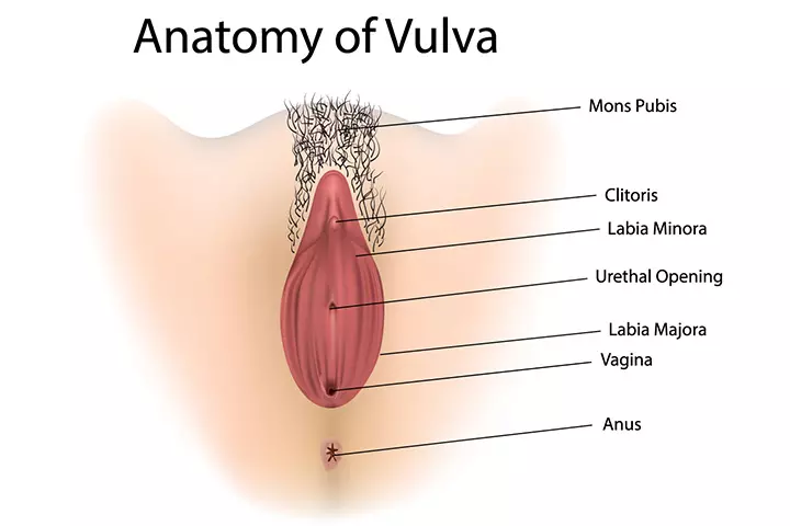
Image: Shutterstock
Vulva is the term for the collective external parts of the female reproductive system. The parts found in the vulva include the mons pubis, the labia, clitoris, and glands, such as Bartholin’s glands and Skene’s glands, which help in lubrication. These parts perform a variety of functions, such as protecting the internal sexual organs, keeping the area moist, and facilitating intercourse.
The following are the various parts of the vulva.
1. Mons pubis: It is a fleshy area located over the pubic bone and above the vagina. Mons pubis is prominent in females and usually covered with pubic hairs after puberty.
Function: Cushioning the pubic bones
2. Labia: These are often referred to as the lips of the vagina and consist of two parts, labia majora and labia minora.
- Labia majora: These are the larger outer prominent pair of lips that cover the labia minora. It is a cutaneous skin fold.
Function: Protection of delicate inner components
- Labia minora: These are a pair of inner smaller lips below the labia majora. It encircles the clitoris and the vulval vestibule.
Function: Protection of clitoris, urethra, and the vaginal opening
3. Clitoris: It is a sensory sexual organ similar to the penis in males. Clitoris is made up of erectile tissues, which cause it to become erect and swollen during sexual arousal. Vestibular bulbs are two band-like tissues related to the clitoris and present on either side of it.
Function: Acts as a pleasure center
4. Vulval vestibule: The openings to the urethra and the vagina are contained in the vulval vestibule, which is outlined by the labia minora. The edge of the vulval vestibule is also referred to as the Hart’s line.
Function: Protects the urethral and vaginal openings
5. Bartholin’s glands: These are also known as the greater vestibular glands and are located posterior to the vaginal opening, one each on either side of the vagina. The pair of glands produce a secretion, which lubricates the vagina during sexual arousal. The secretion also moisturizes the vulva (4). The glands are homologous to bulbourethral glands found in males.
Function: Secretion of a lubricating fluid
6. Skene’s glands: These are also known as the lesser vestibular glands and are located on either side of the urethra. These glands secrete fluid to lubricate the urethra opening and the vulva during sexual arousal.
Function: Secretion of a lubricating fluid
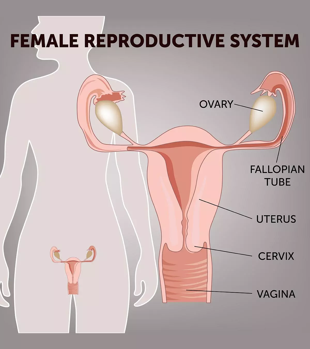
Below are the internal parts of the female reproductive system and their functions.
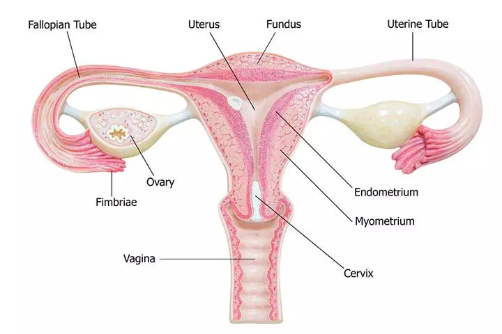
Image: Shutterstock
1. Vagina: It is a muscular canal, anteriorly connected to the vulva, and posteriorly connected to the cervix. The vaginal opening is surrounded by a membrane called the hymen.
Function: It functions as the recipient of the male penis and the sperms (male gametes) during sexual intercourse. The vagina acts as a discharge tract for menstrual flow and also functions as the birth canal for the newborn during vaginal birth.
2. Uterus: It is a pear-shaped organ, also known as the womb, and is located in the pelvis between the bladder anteriorly and the rectum posteriorly. Once an egg or ovum is fertilized, it implants into the inner lining of the uterus. The fertilized egg forms an embryo and then a fetus.
Function: The uterus protects and nourishes the fetus through a specialized tissue called the placenta. The fetus stays in the uterus throughout the pregnancy (gestation), approximately nine months, until it is ready to be delivered.
 Did you know?
The uterus consists of the following parts (5).
- Cervix: It is also known as the neck of the uterus and is part of the uterus that lies between the vagina and the corpus (body) of the uterus.
- Cervical canal: It is a canal that runs through the cervix.
- Corpus: This is the central body of the uterus. It is located behind the cervix and below the fallopian tube openings. Corpus is the part of the uterus where a fertilized egg implants to form an embryo and later develop into a fetus.
- Fundus: This is the uppermost rounded portion of the uterus and lies opposite the cervix.
The thick middle muscle layer of the corpus or the fundus is known as myometrium which expands during pregnancy to hold the baby. The smooth outer layer that covers the uterus is called the serosa or perimetrium .
3. Fallopian tubes: These are also known as the uterine tubes or oviducts and are long slender tubes that connect the ovaries to the uterus.
Function: The ovum (egg) passes from the ovaries to the uterus through the fallopian tubes. Fertilization of the egg usually takes place in these fallopian tubes.
4. Ovaries: These are the primary reproductive organs in the female body. There are two ovaries, one on either side of the uterus (6). Ovaries have an ovoid shape.
Functions: Ovaries are responsible for the productions of the ovum, which is the female gamete. It contains several structures called follicles, which produce an egg that matures during each menstrual cycle. The ovaries also secrete essential hormones, such as estrogen and progesterone.
5. Fimbriae: These are finger-like structures that catch or collect the ovum when released by the ovaries and transfer it to the fallopian tube with a pushing motion.
Function: Unlike sperms (male gametes), the ovum is non-motile, and the fimbriae help provide the necessary motility to the ovum by pushing it towards the uterus.
What Is The Menstrual Cycle?
The menstrual cycle, also called periods, is a 28-day cycle of hormonal changes, uterine tissue growth, tissue breakdown, and tissue removal that occurs in a woman’s body. The menstrual cycle is counted from the first day of a period to the first day of the next period (7).
 Experts say
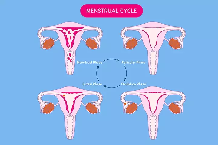
Image: Shutterstock
The following are some salient events of the menstrual cycle.
- The secretion of estrogen and progesterone prepares the uterus for the implantation of a fertilized egg. The endometrium layer, a layer of cells inside the corpus of the uterus, begins to thicken and develop a rich supply of blood vessels.
- If implantation of the fertilized egg does not occur, the levels of estrogen and progesterone start falling. It indicates to the body that pregnancy has not happened.
- The decline in the levels of hormone signals the uterus to discard the upper layer of the endometrium, along with the mucosal tissue. The discarded tissue flows through the cervix and then through the vagina as red blood cell-rich fluid known as menses.
A menstrual cycle is typically 28 days long. However, every woman’s body is different, and so is their menstrual cycle. It is considered a normal menstrual cycle if the periods come every 24 to 38 days.
Below are the key events and their day of occurrence in a typical 28-day menstrual cycle.
- Day 1: First day of the period.
- Day 1 to 7: Follicles develop in the ovary and prepare for the secretion of an ovum.
- Day 8: Menstruation stops. The estrogen and progesterone levels rise to prepare a new lining inside the uterus.
- Day 14: The uterus lining is ready for pregnancy implantation. Ovaries release the mature egg, and ovulation occurs.
- Day 15 to 24: The egg travels through the fallopian tube to the uterus. The sperm meets the egg in the fallopian tube and the embryo is created. Simultaneously, the uterine lining thickens further.
- Day 24 to 28: If fertilization does not occur the unfertilized egg reaches the uterus, and it disintegrates. Therefore, the estrogen and progesterone levels decline, and the excess blood-rich uterine lining sheds and flows out of the vagina as menses or periods.
The cycle is marked by the fluctuation of various hormones, which may affect mood and temperament.
The menstrual cycle begins at puberty and ends at menopause. Menopause usually occurs around the age of 50 years and is a natural process that marks a decline in the ovarian reserve due to depletion of eggs and hence the reproductive hormones.
Functions Of The Female Reproductive System
The following are the key functions of the female reproductive system (8).
- Production of the egg: This process is called oogenesis, in which a female gamete (egg) is produced in the follicles of the ovary. A fertilized egg (embryo) eventually develops into a fetus.
- Ovulation: This is the process of releasing the egg from the ovaries.
- Uterine preparation: The uterus prepares for the possible implantation of the fertilized egg every month.
- Pregnancy: It is also known as gestation and is the process of nourishing the fetus till birth.
- Childbirth: It is also known as parturition and is the process of giving birth to a baby.
- Production of female sex hormones: The ovaries produce estrogen and progesterone hormones, which are called female sex hormones.
20 Facts About The Female Reproductive System
Here are some interesting facts about the female reproductive system (9) (10).
- The ovum is the largest cell of a female body.
- The ovum is about 30 times larger than a sperm cell.
- The female ovaries contain about 300,000 eggs at puberty, which goes on declining as the age progresses. In a lifetime, roughly only 300–400 eggs are ovulated before menopause.
- The lifespan of the ovum is 12–24 hours after it is released from the ovary.
- The zygote is a single-cell entity formed by the fusion of the ovum and the sperm.
- A normal uterus is about three inches long and two inches wide but expands by several times during pregnancy to almost 500 times its size.
- The muscles of the uterus are one of the strongest in the body since they need to contract to push the baby outwards during childbirth.
- The pH of the vagina is acidic and is usually between 3.8 and 4.5 on the pH scale.
- There is no correlation between the hymen “being intact” and virginity, unlike popularly believed. This hymen can rupture during strenuous exercise like horse riding etc.
- A pregnancy can’t be confirmed until the embryo implants on the uterus wall. This is approximately 10-14 days after ovulation and can be confirmed by doing a blood test or urine pregnancy test.
- The menstrual cycle of a woman is usually the most regular during her 20s and 30s.
- The fallopian tube is as wide as a piece of noodle or spaghetti.
- Ovaries are part of the reproductive system and the endocrine system since they secrete hormones, too.
- A woman can get pregnant during her periods, too, if the other ovary ovulates at that time. Although the occurrence of this is quite rare.
- Each ovary is about the size of an almond.
- There are tiny hair-like structures inside the fallopian tube that gently sway and push the egg towards the uterus. These are called cilia.
- The cancer of the ovaries is called ovarian cancer, which is hard to detect in the early stages since it may cause no symptoms or mild symptoms.
 Quick fact
- Many women develop ovarian cysts, which are fluid-filled pockets on ovaries. This is known as polycystic ovarian disease (PCOD), which usually needs treatment.
- Some women may have such regular periods that they may predict the precise day and time for the periods to begin.
Frequently Asked Questions
1. How big is the female reproductive system?
The uterus is around 5cm wide by 7cm long in a non-pregnant woman.
The vagina is around 10cm long. The ovaries are around 2 to 3cm long (11).
2. How much does the uterus weigh?
The uterus weighs around 30 to 40 grams (12).
3. What are the common diseases and conditions associated with the female reproductive system?
The female reproductive system can be affected by various diseases and conditions. Some common health concerns include polycystic ovary syndrome (PCOS), endometriosis, uterine fibroids, interstitial cystitis, and sexually transmitted infections. Other conditions are gynecological ovarian, cervical, or uterine cancers. Therefore, seeking the advice of a healthcare professional is essential for the accurate diagnosis, treatment, and management of these disorders (16).
4. How does the female reproductive system change throughout life?
A woman’s reproductive system undergoes significant changes during her lifetime. During puberty, the reproductive organs develop, and menstruation begins. In the reproductive years, the system is primed for fertility and conception. As a woman approaches menopause, hormone levels fluctuate, leading to the cessation of menstruation and the end of fertility. Additionally, aging can result in changes to the reproductive organs, such as decreased ovarian function and alterations in the uterine lining. These changes reflect the natural progression of a woman’s reproductive system from puberty to menopause (17).
5. What can I do to maintain reproductive health?
To keep your reproductive system healthy, eat a balanced diet with plenty of vitamins and minerals and avoid sugary, processed foods and unhealthy fats. Instead, choose fresh fruits, vegetables, whole grains, and foods high in fiber and omega-3s. Practice safe sex to prevent STIs, which can affect your reproductive health. Also, maintaining a healthy weight and managing stress are essential.
The female reproductive system is a complex system that does the wonder of procreation. It is important for children to learn about the different parts of this system to understand how a female body functions. Learning the facts about this system is the first step toward sex education for children. Different parts of the female reproductive system, menstrual cycle, and other functions of the system may help your children develop an interest in biology and science. In addition, it will also help young girls and boys understand how pregnancy occurs and, therefore, learn the importance of safe sex.
Infographic: Facts About The Female Reproductive System
The female reproductive system is a complex and vital system that plays a key role in reproduction and overall health. This infographic provides important information about its functions, from the ovaries to the uterus. Read along and share this knowledge with others as well.
Some thing wrong with infographic shortcode. please verify shortcode syntax
Prepare to uncover the secrets of the organs that comprise the female reproductive system. In this informative video, we will delve into the fascinating intricacies of the female reproductive system.
References
1. Reproductive Anatomy and Physiology; Winchester Hospital
2. Female Reproductive System; Cleveland Clinic
3. John Nguyen and Hieu Duong, Anatomy, Abdomen and Pelvis, Female External Genitalia; NCBI
4. Min Y. Lee et al., Clinical Pathology of Bartholin’s Glands: A Review of the Literature; NCBI
5. Hilary O. D. Critchley, Physiology of the Endometrium and Regulation of Menstruation; American Physiological Society
6. Ovaries; National Cancer Institute
7. Your menstrual cycle; U.S. Department of Health & Human Services
8. Suzanne Wakim and Mandeep Grewal, Functions of the Female Reproductive System; LibreTexts
9. What is the strongest muscle in the human body?; Library of Congress
10. Female Reproductive System; Rady Children’s Hospital, San Diego
11. Anatomy and Physiology of the Female Reproductive System; Openstax
12. UTERUS; King George’s Medical University
13. Fun Facts About the Placenta; Oregon Health & Science University.
14. About Heavy Menstrual Bleeding; CDC
15. Ovarian Cancer Statistics; CDC.
16. Common Reproductive Health Concerns for Women; CDC
17. Aging changes in the female reproductive system; Medline Plus
Community Experiences
Join the conversation and become a part of our nurturing community! Share your stories, experiences, and insights to connect with fellow parents.
Read full bio of Dr. Rana Choudhary (Khan)
Read full bio of Dr Bisny T. Joseph
Read full bio of Harshita Makvana
Read full bio of Shinta Liz Sunny


