Image: ShutterStock
A growth scan during pregnancy is carried out to check the baby’s development. Often scans and screening tests are a part of routine care in pregnancy and are conducted before the third trimester. Growth scans are typically performed in the final trimester for most women. Nevertheless, it may cause you to become concerned and anxious about your baby’s health.
Read on to know more about growth scans and understand their significance in pregnancy.
Key Pointers
- A growth scan is an ultrasound scan done in the third pregnancy trimester to ascertain the proper growth and development of the fetus.
- A growth scan evaluates the overall growth of the baby, level of amniotic fluid, position and maturity of the placenta, and fetal position and weight.
- Doctors may suggest growth scans for reasons such as a history of pregnancy-related complications, pre-existing medical conditions, decreased fetal movements, and multiple pregnancies.
- Doctors use the results of the growth scan to determine any potential risks, fetal abnormalities, and also to decide the time and method of delivering the baby.
What Is A Growth Scan During Pregnancy?

Image: Shutterstock
If you have a scan during your third trimester, it’s called a ‘growth scan’ or ‘wellbeing scan’ – which is done to determine your baby’s size and measurements and assess their health. A growth scan is an ultrasound scan that determines whether your baby’s growth is normal. Doctors typically recommend it for women during the third trimester of pregnancy; one of the main reasons is to know about fetal growth (1).
What Is The Purpose Of A Growth Scan?

Image: Shutterstock
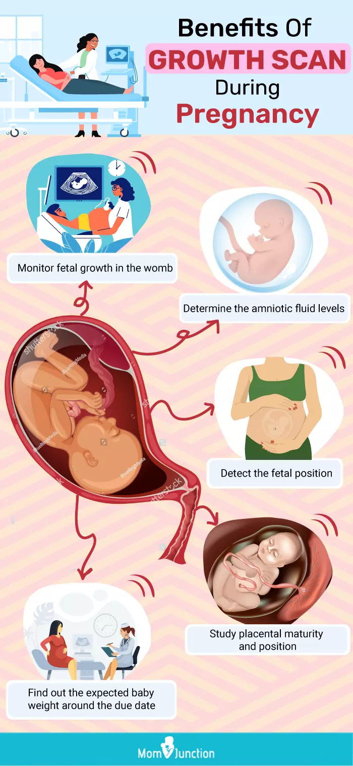
A growth scan in obstetrics during pregnancy, which is also commonly known as a sonogram, is performed to provide your doctor with useful information about your pregnancy and fetal growth. It helps the doctor (2):
- Check your baby’s overall growth.
- Check the volume of your amniotic fluid.
- Determine the final position and maturity of the placenta.
- Determine the fetal position.
- Check the baby’s estimated weight close to your delivery date [3].
A mom and blogger, Rebecca, shares how a growth scan eased her doctor’s concerns about her baby’s growth. She says, “As I am quite tall and my body contained everything compactly, by 28 weeks, my OB was concerned about the baby’s growth, and I had extra growth scans… but as it turned out, he was just a petite baby with long legs like his parents! (i)”
Specific measurements from the growth scan can be used to estimate fetal growth. A fetal growth chart serves as a reference to assess the growth of the fetus. The below graph shows the estimated fetal weight with gestational age. The lines represent different growth rates, and the red one denotes the average growth rate or the 50th percentile. The lowest and highest growth is represented by the black top and bottom lines, which correspond to 99% and 1%, respectively.

Estimated fetal weight by gestational age
Source: The World Health Organization fetal growth charts: concept, findings, interpretation, and application; American Journal of Obstetrics and Gynecology/WHOWho Needs A Growth Scan?
You may need a growth scan for various reasons. Some of these reasons include (3):
- If you have had complications in your previous pregnancies, your doctor may perform additional scans to monitor the fetal health. Pregnancy complications can mean health risks for you and the fetus. If you had a medical problem like thyroid disease, seizures, asthma, heart disease, or anemiaiA condition characterized by reduced levels of red blood cells and hemoglobin in the body during pregnancy. before or during pregnancy, it can lead to pregnancy complications. Similarly, accidents or injuries while pregnant can also lead to pregnancy complications. In such cases, doctors may opt for a late pregnancy growth scan to keep a tab on the pregnancy complications that can arise due to health problems (4).
- Pregnancy complications can also arise due to the placental position and placental breaks. In the following two conditions (5):
- Placenta previa means the placenta is laying low in the womb. It can block the cervixi The lowest portion of the uterus that connects to the vagina to form a passage between the two organs. and make vaginal delivery difficult or impossible and can lead to severe bleeding.
 Research finds
Image: Shutterstock
- Placental abruption is when the placenta breaks away from the uterine wall due to some health complications and bleeding through vagina is observed.
Your doctor will be monitoring your pregnancy and fetal health frequently for the rest of your term. A growth scan and assessment in the last few weeks of your pregnancy may be essential to determine the placental position and so to decide the time and method of delivering your baby [6].
- Many a time, women develop diabetes during pregnancy (gestational diabetes). It is a serious condition, and if you don’t manage it well, it can cause pregnancy complications. A growth scan helps your doctor determine if there is a problem with your pregnancy due to gestational diabetes. Your baby can suffer from excess birth weight, which may mean a C-section delivery for you. Your child can have respiratory problems, a preterm birth, and low sugar at birth. These are just some of the risks to your baby due to gestational diabetes. Women with a history of gestational diabetes or preterm labor may need frequent monitoring. With the help of a growth scan, your doctor can determine if your pregnancy or baby has any problems due to your gestational diabetes (6).
- Women may develop high blood pressure during pregnancy (gestational hypertension). Whether you suffer from high blood pressure before your pregnancy or gestational hypertension, both conditions can cause pregnancy complications. In case you have high blood pressure during pregnancy, your doctor will like to monitor your baby’s growth regularly. And, a growth scan can help your doctor check for fetal growth abnormalities and complications (7).
- The doctor usually measures the fundal height. Fundal height is the distance from the top of your uterus to your pubic bone. It should correspond with your pregnancy in weeks. For instance, if you are 22 weeks pregnant then your ideal fundal measurement is 22 cm.
If there is a discrepancy in this measurement, it can indicate
- slow or rapid fetal growth.
- too little or excess amniotic fluid,
- Change in fetal positions other than normal or
- uterine fibroidsiBenign uterine growths that may cause heavy and prolonged menstrual bleeding and back pain. .
So a growth scan may then be necessary to make a diagnosis and find out the problems with fetal health (8).
- Your fundal height can also indicate fetal growth restriction or intrauterine growth restriction (IUGR); both indicate low fetal weight or abnormal fetal growth rate. Your doctor may then confirm it through a pregnancy ultrasound. If the ultrasound results indicate that the fetal growth is not optimum, your doctor is likely to perform a growth scan (or more than one) as your pregnancy progresses towards the end of your term (8).
- Multiple pregnancies can lead to more risks or complications. A growth scan in the third trimester of your pregnancy can help the doctor ascertain if there is any cause for concern (9).

Image: Shutterstock
- You begin to experience less fetal movements during your third trimester. But If it persists, your doctor may perform a growth scan to check if everything is alright with your baby (10).
- If you have a fetal breech position. Breech means fetal bottom-down position. It is not an ideal birth position, and you may need to undergo a C-section. If an earlier ultrasound scan revealed the breech position, your doctor might recommend another growth scan towards the end of your pregnancy to check the position of the fetus and the placenta (11).
 Quick fact
Your doctor could suggest a growth scan during pregnancy to check your health and fetal development. It is a vital ultrasound scan generally performed in the third trimester to assess your amniotic fluid volume, placental position, the baby’s height, weight, and overall growth. However, women who have pregnancy-related complications may need growth scans more frequently to monitor the fetus. Altogether, the scan is helpful to detect any anomalies that help your doctor suggest prompt, necessary steps to enable a safe delivery with a healthy baby.
Frequently Asked Questions
1. What happens if a growth scan shows a big baby?
A growth scan showing a big baby could mean that the baby is large for gestational age (LGA). Diabetes in mothers or genetics could be the cause of LGA. If the baby is diagnosed with LGA, the healthcare provider may recommend an early delivery to prevent complications (12).
2. How long does a growth scan take?
The growth scan is a simple, non-invasive procedure performed by a professional sonographer or a doctor. The scan takes approximately 30 minutes to study and determine the fetus’ health.
No. A growth scan is a precautionary measure to ensure that your fetus is growing well without complications. In a normal pregnancy, you may be asked for a growth scan around the 34th week of pregnancy; however, in a high-risk pregnancy, it may be suggested around the 28th week (1).
3. How often are growth scans typically done during pregnancy?
Depending on the risk associated with your pregnancy, you will be advised additional growth scans during the third trimester. For a high-risk pregnancy, there will be five scans at 28, 31, 34, 37, and 40 weeks of gestation. In the case of a moderate-risk pregnancy, scans will be offered at 32, 36, and 40 weeks. If your pregnancy is classified as moderate-low risk, two scans will be scheduled at 32 and 36 weeks (14).
4. What potential causes for slow growth are detected during a growth scan?
Several maternal factors can contribute to fetal growth restriction (FGR). These include high blood pressure or other heart and blood vessel diseases, diabetes, anemia, long-term lung or kidney conditions, autoimmune conditions like lupus, being underweight or overweight, poor nutrition or weight gain, alcohol or drug use, and cigarette smoking. On the other hand, factors in the baby that can lead to FGR include being one of multiple pregnancies (twins or triplets), infections, birth defects such as heart defects, and problems with genes or chromosomes. These various factors can hinder the baby’s normal growth and development during pregnancy (15).
5. What are the alternatives to growth scans for monitoring fetal growth?
A Doppler ultrasound can also be taken from the regular ultrasound to monitor fetal growth. Doppler sonography is a technique that checks the blood flow in the baby to see if it’s getting enough oxygen. It does this by measuring the resistance to blood flow and looking at the pattern of blood movement during the heartbeat. It also measures how fast the blood is moving in the vessels. If there are any abnormalities in the blood flow, it can indicate problems with the baby’s circulation. Many studies have shown that using Doppler measurements is helpful in managing babies who are not growing well in the womb. It helps doctors ensure the pregnancy continues safely, reducing the risk of premature birth, stillbirth, or long-term effects on the baby’s health (16).
Infographic: What Is The Purpose Of A Growth Scan?
When pregnant, you may be advised to have a growth scan during the third trimester to track your baby’s growth. However, a doctor may order it more than once if you have any pregnancy-related complications. If you are interested in learning the objectives and advantages of growth scans, this infographic is a worthy read. Illustration: Momjunction Design Team
Illustration: Growth Scan During Pregnancy – Everything You Need To Know

Image: Stable Diffusion/MomJunction Design Team
You may wonder about the safety of growth scans and their accuracy. Get your questions answered in this video! Learn if you need a growth scan and how accurate they actually are.
Personal Experience: Source
MomJunction articles include first-hand experiences to provide you with better insights through real-life narratives. Here are the sources of personal accounts referenced in this article.
i. Pregnancy with Rowan.https://mypregnancytruths.blogspot.com/p/pregnancy-with-rowan.html
References
- Growth Scan.
https://www.fetalclinic.org/growth-scan-2/ - Harold Gomez Acevedo et al.; 2022; Sonography 3rd Trimester And Placenta Assessment, Protocols, And Interpretation.
https://www.ncbi.nlm.nih.gov/books/NBK572062/#:~:text=The%20use%20of%20ultrasound%20in - Ultrasound pregnancy.
https://medlineplus.gov/ency/article/003778.htm - Diann M. Krywko; Pregnancy Trauma.
https://www.ncbi.nlm.nih.gov/books/NBK430926/ - What complications can affect the placenta?
https://www.nhs.uk/pregnancy/labour-and-birth/what-happens/placenta-complications/ - Gestational Diabetes.
https://www.acog.org/womens-health/faqs/gestational-diabetes - High Blood Pressure in Pregnancy.
https://medlineplus.gov/highbloodpressureinpregnancy.html - Fundal Height.
https://my.clevelandclinic.org/health/diagnostics/22294-fundal-height - Multiple pregnancy: having more than one baby.
https://www.rcog.org.uk/for-the-public/browse-our-patient-information/multiple-pregnancy-having-more-than-one-baby/ - Prenatal care in your third trimester.
https://medlineplus.gov/ency/patientinstructions/000558.htm - Breech birth.
https://medlineplus.gov/ency/patientinstructions/000623.htm - Large for gestational age.
https://www.stanfordchildrens.org/en/topic/default?id=large-for-gestational-age-90-P02383 - 22+ Weeks Scan (Growth & Wellbeing Scan).
https://womensimaging.net.au/what-we-do/obstetrics/growth-wellbeing-scan/ - 3rd Trimester Obstetric Ultrasound Scans Fetal Growth Assessment;
https://www.hey.nhs.uk/patient-leaflet/3rd-trimester-obstetric-ultrasound-scans-fetal-growth-assessment/ - Fetal Growth Restriction;
https://www.urmc.rochester.edu/encyclopedia/content?ContentTypeID=90&ContentID=P02462 - Zarko Alfirevic; Fetal and umbilical Doppler ultrasound in normal pregnancy.
https://www.ncbi.nlm.nih.gov/pmc/articles/PMC6464774/#:~:text=Doppler%20ultrasound%20uses%20sound%20waves,the%20baby’s%20condition%2C%20shows%20benefits. - Frances M. Anderson-Bagga & Angelica Sze; Placenta Previa.
https://www.ncbi.nlm.nih.gov/books/NBK539818/#:~:text - Caron J. Gray & Meaghan M. Shanahan; Breech Presentation.
https://www.ncbi.nlm.nih.gov/books/NBK448063/#:~:text=
- Growth Scan.
Community Experiences
Join the conversation and become a part of our nurturing community! Share your stories, experiences, and insights to connect with fellow parents.
Read full bio of Dr. Anita Gupta
Read full bio of shreeja pillai
Read full bio of Rebecca Malachi
Read full bio of Reshmi Das


